

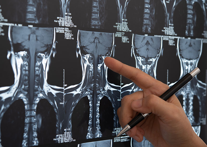
Cervical disc fusion is a surgery that fuses together selected bones in the neck (cervical spine). It is most often performed to treat conditions of the spine such as spinal stenosis, a herniated disc, osteoarthritis, infections, tumours, spine deformities, and misalignments of the vertebrae. It can also be performed after an injury to stabilize the neck and prevent a bone fracture from causing instability or damage.
Depending on the individual’s circumstances, there are different methods of performing a cervical disc fusion, and the procedure can be performed through an incision on the front (anterior) or back (posterior) of the neck, as detailed below.
Dr. Cochrane performs this surgery using a minimally invasive technique where he makes a small incision on the front (anterior) of the neck, which gives him direct access to the disc through a relatively uncomplicated pathway.
The surgery itself is made up of two parts:
1. Discectomy – First, Dr. Cochrane makes an incision through the front of the neck and removes the damaged disc.
2. Fusion – He then inserts a bone graft or an implant device in the empty space where the damaged disc was to provide strength and stability to the area.
The procedure may also involve removing part of the vertebra or widening the spinal canal to give the spinal cord more space if neurological symptoms from spinal cord compression are present.
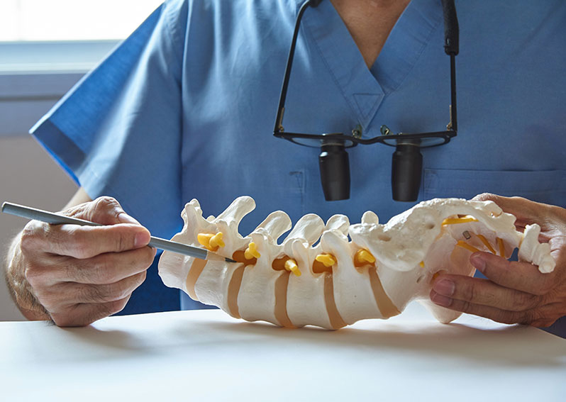
Posterior cervical fusion is the general term used to describe the procedure of mending two or more cervical vertebrae through an incision in the back of the neck.
During a posterior cervical fusion, Dr. Cochrane makes an incision in the back of the neck and places a bone graft along the sides of the injured section of the cervical spine. Over time, this bone graft fuses together to provide healing and greater stability.
Metal screws and rods are sometimes used in conjunction with the procedure to extend fusion and/or provide immediate stability and increase the likelihood of successful fusion.
During this procedure, Dr. Cochrane removes the injured disc and replaces it with an artificial disc, which is designed to replicate the natural motion of the cervical spine in order to preserve range of motion in that area of the neck. The artificial disc core slides and rotates inside the disc, self-adjusting to the patient’s cervical spine’s movements, which means it can react to the normal motion in the cervical spine.
Dr. Cochrane typically performs this surgery for patients who have cervical disc herniation or cervical degenerative disc disease that causes chronic neck and/or arm pain that hasn’t been relieved from non-surgical treatments.
This type of surgery, allows you to:
The procedure is designed to relieve the pressure on your spinal cord or nerves that are causing the pain, numbness, and weakness that can radiate to your shoulder, arm, and hand.
Dr. Cochrane performs the cervical total disc replacement through a small incision on the neck. After he removes the damaged disc, he will smooth away any bone spurs and then place and anchor the artificial disc in the empty space that is left. Dr. Cochrane then closes the incision and covers it with a dressing.
You will then be moved to a recovery room where you will be monitored for the next few hours. After you have recovered, you will be sent home with post-op instructions. Dr. Cochrane may also recommend you wear a soft collar for a short period of time to add support to your neck.

Dr. Cochrane performs cervical decompression surgery to relieve compression of one or more of the vertebrae located in your neck. This compression can occur for a number of different reasons. One of the most common is the gradual wear and tear on the cervical spine that typically occurs in people over 50 years of age.
Conditions that cause cervical compression usually develop more quickly and occur at any age. These include:
Cervical decompression surgery is a procedure designed to remove structures such as bone spurs that are compressing the nerves in your neck while widening the space between vertebrae. Removal of these small structures should alleviate pressure and allow the nerve to heal.
The goal of cervical decompression surgery is to leave the cervical spine intact. However, in some situations, there are so many bony structures pressing on the nerve that removing them may cause instability. If this is the case, cervical decompression surgery Dr. Cochrane may also perform cervical fusion surgery to correct the instability.
Anterior lumbar interbody fusion (ALIF) is one of the most frequently used spinal fusion techniques and is performed through the front of the body (anterior approach). Interbody fusion refers to the removal of an intervertebral disc, which is replaced with a spacer during the fusion process. A major advantage of anterior entry is that a larger implant can be incorporated into the procedure.
Dr. Cochrane typically performs the ALIF procedure to treat deformity of the spine, nerve compression, and associated pain. Such compression of spinal nerves may occur as a result of progressive disc degeneration, slippage of the vertebrae (spondylolisthesis), abnormal curvature of the spine (scoliosis, kyphosis, or loss of lordosis), or other causes of progressive spinal instability. The ALIF procedure is typically used to fuse either the lowest level of the spine (L5/S1), the second-lowest level of the spine (L4/5), or both.
Patients with persistent low back pain, which often radiates down the leg, may be candidates for ALIF if more conservative treatments, such as rest, non-steroidal anti-inflammatories (NSAIDs), physical therapy, and corticosteroid injections, have not been effective in relieving their symptoms.
Patients undergo the ALIF procedure under general anesthesia and lying face-up on an operating table. The surgeon makes a small incision on the side of the abdomen near the affected area through which the injured disc and other debris are removed.
A cage (or spacer) is then inserted in the disc space. The cage is typically made of either titanium or plastic and packed with bone graft. After inserting the cage, the surgeon will place either a metal plate or screws and rods in the vertebrae to maintain stability of the spine around the cage as the vertebrae grow together and fuse.
After undergoing an ALIF procedure, patients typically remain in the hospital overnight. Sometimes, a longer length of stay is required if the ALIF is performed as part of a multi-stage procedure. A physical therapy regimen is started soon after to assist the patient in regaining strength and mobility. Certain activities may be restricted, including lifting, twisting the midsection, and bending at the waist. Many patients can return to work within a few weeks after the procedure if their employment does not require strenuous exertion. However, heavy lifting and manual labor are prohibited for 3-6 months after the procedure, depending on the extent of the surgery.

The lumbar spine, or lower back, consists of five vertebral bones lined one above the other and separated by spongy gel-like discs that cushion the spine during movement. Over the years, these discs can undergo wear and tear, which can cause the vertebrae to painfully rub against each other. In turn, this movement can lead to degeneration, which then leads to lower back pain as well as pain, numbness, or weakness in the legs.
Oblique lumbar interbody fusion (OLIF) is a minimally invasive procedure that involves removing the damaged intervertebral disc and bone then fusing together the two adjacent vertebrae.
If a patient is not responding to conservative treatments and quality of life is affected, then OLIF surgery may be the next step. OLIF may be recommended for patients suffering back pain due to degenerative disc disease, disc herniation, spinal stenosis, spondylolisthesis, scoliosis, fractures , and spine instability.
During OLIF surgery, Dr. Cochrane makes a small lateral incision in the side of the abdomen to spare the disruption of major back muscles, ligaments and bones, and preserves back strength. The damaged vertebral bone or intervertebral disc is partially or totally removed. Bone graft or a suitable spacer is placed to maintain the intervertebral space and allow the bones to fuse. Screws, rods and plates may be used for additional support. The soft tissues are carefully repositioned and the incision closed.